

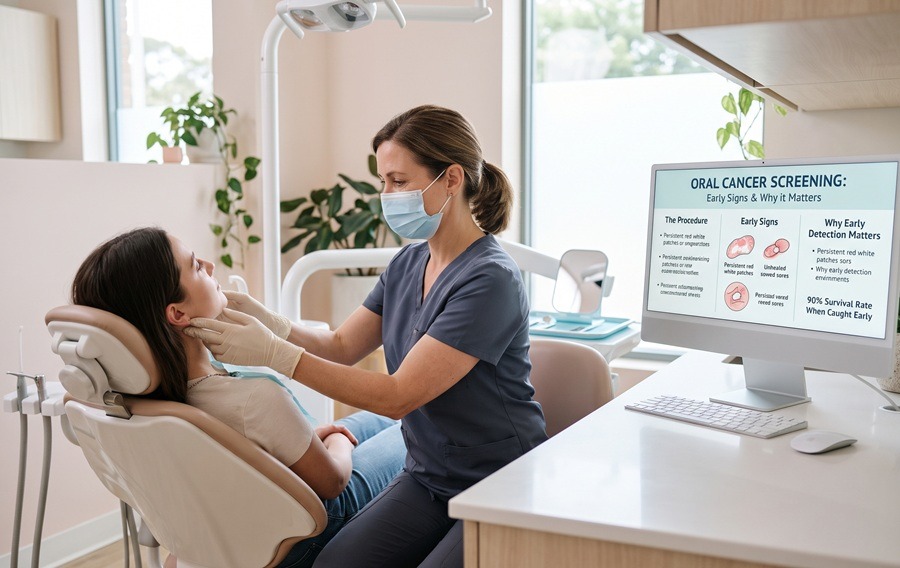
An oral cancer screening is a brief, painless clinical examination performed by a dentist or periodontist to detect precancerous lesions or early-stage signs of cancer in the oral cavity and throat before physical symptoms appear. By combining visual tissue inspections, gentle head and neck palpation, and optical technology, a routine mouth cancer screening identifies abnormal cellular changes when treatment is simplest and survival rates exceed 80% to 90%.
Most people associate dental visits strictly with teeth and gums, rarely thinking about the delicate soft tissues lining their cheeks, tongue, and palate. Yet oral cavity and oropharyngeal cancer often develops silently, without pain or obvious discomfort in its earliest stages. By the time a lesion hurts or interferes with swallowing, it has frequently progressed into deeper tissue layers.
At Golden State Dental Co., our board-certified periodontal and surgical specialists integrate thorough soft-tissue evaluations into every diagnostic visit across our Sacramento, Folsom, and Roseville offices. Understanding how an oral cancer examination works empowers you to stay proactive about both your oral and systemic wellness.
What is an Oral Cancer Screening? (Purpose and Goals)
The primary goal of an oral cancer screening is to catch mucosal abnormalities—such as persistent red or white patches, tissue thickening, or subtle ulcerations, at their earliest, most curable stage.
Your mouth is lined with specialized skin called mucosa, which naturally renews itself every 10 to 14 days. When cells along the tongue, floor of the mouth, gums, or tonsillar pillars begin dividing abnormally due to genetic mutations, chronic irritation, or viral factors, they leave subtle surface clues long before a tumor forms.
Specifically, clinicians look for two classic precancerous indicators during a screening:
- Leukoplakia: Thickened, firmly attached white or grayish patches inside the mouth that cannot be scraped away. While most leukoplakia is benign irritation, a percentage shows dysplasia (precancerous cellular changes).
- Erythroplakia: Flat or slightly raised velvety red patches that often bleed easily when touched. Erythroplakia carries a significantly higher risk of harboring early malignancy and requires prompt evaluation.
What to Expect During the Screening: The Step-by-Step Procedure
A complete oral cancer examination takes less than five minutes, requires no needles or downtime, and is seamlessly built into your comprehensive periodontal checkup.
Step 1: Visual Inspection (Inside the Mouth)
Your specialist uses a bright diagnostic light and a dental mirror to systematically examine every surface of your oral cavity:
- The lips, inner cheeks (buccal mucosa), and gum tissue (gingiva)
- The roof of the mouth (hard and soft palate) and back of the throat (oropharynx and tonsils)
- The top, underside, and lateral borders of your tongue—the most common site for intraoral lesions
- The floor of the mouth beneath your tongue, gently holding the tip of your tongue with gauze to get a clear view
Step 2: Physical Palpation (Head and Neck)
Not all abnormalities are visible on the surface. Using gloved fingertips, your doctor gently presses along the floor of your mouth, your jawline, beneath your chin, and down the sides of your neck to check your lymph nodes and salivary glands. They are feeling for firm, fixed nodules, asymmetrical swelling, or tissue induration (hardening beneath the surface).
Step 3: Adjunctive Screening Technologies (Specialized Aids)
When a clinician wants a closer look at questionable tissue or wishes to screen high-risk patients with greater sensitivity, specialized clinical aids can assist the visual exam:
- Fluorescence Optical Technology (VELscope): A handheld device that emits a safe blue-spectrum light into the mouth. Healthy soft tissue naturally fluoresces a bright apple-green pattern, while abnormal, dysplastic, or highly vascular tissue absorbs the light and appears distinctly dark.
- Toluidine Blue Dye Screening: A specialized vital dye applied with a cotton swab to a suspicious mucosal spot. Because precancerous and malignant cells contain higher concentrations of nucleic acids and divide rapidly, they retain the dark blue stain after rinsing, helping pinpoint the exact area that needs a biopsy.
- Brush Cytology: A painless soft brush used to collect surface cells from a minor lesion for microscopic laboratory analysis.
How to Prepare for an Oral Cancer Screening?
No fasting, medication adjustments, or special preparation is required before an oral cancer screening.
To help your clinician get the clearest view of your oral tissues:
- Remove Dental Appliances: If you wear removable partial dentures, full dentures, or orthodontic retainers, you will be asked to take them out so the underlying gums and palate can be inspected completely.
- Share Your Symptom History: Let your doctor know if you have noticed a canker sore that hasn’t healed after two weeks, chronic hoarseness, or a persistent feeling that something is caught in the back of your throat.
- Skip Heavy Colored Foods Right Before Your Visit: Avoid hard candies, berries, or colored sports drinks immediately prior to your appointment so artificial dyes don’t temporarily stain your tongue or mucosa.
Understanding the Limitations of Oral Cancer Screening
While routine screenings are a vital frontline defense, transparency about oral cancer screening limitations helps patients understand what the exam can and cannot do:
| Clinical Factor | What a Screening Does | The Limitation to Keep in Mind |
| Screening is Not a Diagnosis | Flags abnormal tissue color, texture, or firmness | Cannot confirm cancer visually; only a laboratory biopsy provides a definitive diagnosis |
| False Positives | Catches every atypical spot, including benign irritation | Cheek biting, hot food burns, or lichen planus can mimic precancerous patches and cause temporary worry |
| Hidden Areas | Thoroughly evaluates the visible oral cavity and neck | Deep HPV-related oral cancer tucked inside the base of the tongue or tonsil crypts can be hidden from plain sight |
Early Warning Signs and Symptoms You Should Never Ignore
Because everyday mouth sores (like a bitten cheek or minor aphthous ulcer) heal within 10 to 14 days, any abnormality that lingers past the two-week mark warrants a professional look. Watch for:
- A sore, ulcer, or blister on the lip, tongue, or gums that does not heal within 14 days
- Persistent white (leukoplakia), red (erythroplakia), or speckled red-and-white patches
- A painless lump, thickening, or rough crusty spot inside the cheek or along the neck
- Unexplained numbness, tingling, or loss of sensation in the lower lip, chin, or tongue
- Difficulty chewing, swallowing (dysphagia), or moving the jaw and tongue freely
- Unexplained loosening of one or more teeth without generalized periodontal disease, or dentures that suddenly stop fitting properly
- Chronic sore throat, ear pain on one side, or a persistent change in the pitch of your voice
Major Risk Factors: Who Needs Screenings Most Frequently?
Historically, mouth cancer primarily affected older adults with a history of heavy tobacco and alcohol use. Today, the clinical landscape has shifted dramatically due to viral factors, meaning even young, non-smoking patients require routine screenings:
- Tobacco Use of Any Kind: Cigarettes, cigars, pipes, chewing tobacco, and snuff expose mucosal cells directly to carcinogens.
- Heavy Alcohol Consumption: Alcohol dehydrates the oral mucosa, making tissue significantly more permeable to toxins. Combining tobacco and alcohol multiplies oral cancer risk up to 30-fold compared to abstaining from both.
- HPV-Related Oral Cancer: Infection with Human Papillomavirus (specifically the HPV-16 strain) is now the leading cause of oropharyngeal cancer (affecting the back of the throat, base of the tongue, and tonsils) in the United States—frequently diagnosed in healthy nonsmokers under age 50.
- Chronic Sun Exposure: Prolonged ultraviolet (UV) exposure without lip protection is a primary driver of lower lip cancers, particularly for patients working or exercising outdoors in sunny California.
- Age, History & Immune Suppression: Adults over 45, patients with a prior history of head or neck dysplasia, and individuals with weakened immune systems benefit from more frequent 3-to-6-month periodontal evaluations.
What Happens If an Abnormal Spot or Lump Is Found? (Next Steps)
First, take a deep breath: the vast majority of spots discovered during a mouth cancer screening turn out to be completely harmless. Traumatic bites, sharp tooth edges, minor fungal infections, and benign fibromas are far more common than malignancy.
If your specialist identifies an area of concern, they will follow a clear, structured protocol:
- The 14-Day Re-Evaluation: If the spot looks like trauma or irritation (for example, from a sharp filling or hot food burn), your doctor will eliminate the source of friction, take a clinical photograph with measurements, and have you return in two weeks to confirm it has healed.
- In-Office Biopsy or Biopsy Referral: If a lesion persists beyond two weeks or displays high-risk characteristics right away, a small tissue sample (an incisional or excisional biopsy) is taken under local anesthesia—either right in our surgical suite or via a coordinated biopsy referral to an oral pathology laboratory.
- Pathology Review & Coordinated Care: A board-certified oral pathologist examines the cells under a microscope and delivers a definitive report within days, ensuring you either get complete peace of mind or an immediate, coordinated care plan.
Frequently Asked Questions About Mouth Cancer Screenings
How often should I get an oral cancer screening?
Most adults should receive a professional oral cancer examination at least once every six months during their routine dental or periodontal checkup. Patients who use tobacco, consume alcohol regularly, have a history of oral dysplasia, or manage chronic periodontal disease should be screened every 3 to 4 months.
Does an oral cancer screening hurt?
No, an oral cancer screening is completely non-invasive and painless. It involves only a visual look with a bright light and gentle finger pressure along your jaw and neck. Even if adjunctive tools like a VELscope light or toluidine blue dye screening are used, there is zero discomfort.
Can a regular dentist or periodontist diagnose oral cancer on the spot?
No clinician can definitively diagnose cancer by sight or touch alone. While a periodontist or oral surgeon has advanced training to recognize suspicious leukoplakia and erythroplakia, only a microscopic tissue bopsy analyzed by a pathologist can confirm whether abnormal cells are benign, precancerous, or malignant.i
Protect Your Overall Health: Schedule Your Screening at Golden State Dental Co.
Your mouth is a window into your body’s overall health, and five minutes in the dental chair can quite literally save your life. Whether you are due for your routine periodontal maintenance or have noticed a small spot inside your mouth that you want a specialist to evaluate, our team is here to provide clear answers and compassionate care.
Contact Golden State Dental Co. today to book an appointment at our Sacramento, Folsom, or Roseville offices. Let our board-certified specialists safeguard your smile and your long-term health.
Get More from Golden State Dental Co
Add us as a preferred source to see more of our content on Google Search.
